Note: Descriptions are shown in the official language in which they were submitted.
1
SETTING OF MULTIPLE PRIMING OLIGONUCLEOTIDES FOR SOLID GEL
AMPLIFICATION IN HYDROGELS
FIELD OF THE INVENTION
The present invention pertains to the field of macro- and microfluidic devices
and
methods for detection of nucleic acids
BACKGROUND OF THE INVENTION
There is an increasing demand for a small scale array-based and/or
microfluidic device
that processes micro- or nano-volumes of sample, with time and cost savings
arising
from miniaturization. Prior art approaches to miniaturised polymerized chain
reactions
("PCR'') make use of open or enclosed chambers or flow through zones/channel
networks with appropriate temperature regulation; some have on-board silicone
rubber-
based or magnetic-based valving and/or pumping. Although potentially powerful
approaches, challenges may arise of pressure seal and/or evaporation, pressure
buffering, as well as others such as chemical interference through surface
interactions,
and evaporation/contamination via the porous, gas permeable membranes used in
pumps and valves.
Performing PCR in a colloidal hydrogel matrix (hereafter termed "gel") may
confer a
multitude of advantages. For example, the DNA, polymerase enzyme and other PCR
reagents a) have reduced access to the device materials' surfaces where they
may be
adsorbed, absorbed, poisoned or otherwise rendered inactive and b) are kept
within
close proximity to each other without the need for valves or pumps. Likewise,
any
contaminant solutes from device materials have reduced access to the PCR
reaction.
Gels provide a successful medium for PCR, as first introduced by Chetverin et
al., see
for example U.S. Pat. No. 5,616,478. PCR was confined to circular spots in a
gel sheet
where the initial DNA or RNA templates, formed "molecular polonies' (short for
CA 2809729 2017-12-14
2
polymerase colonies), named for their similarity to the growth of bacterial
colonies in
agar; the initial amount of DNA can be accurately estimated by counting the
number of
polonies. Mitra et al. (Mitra, R. D. et al; Nucleic Acid Research 1999, 27,
e34)
performed DNA amplification in a thin acrylamide film polymerized with all the
reagents along with plasmid DNA as their template. In an alternate approach,
Strizhkov
et al. (Strizhkov, B. N. Et al; Biotechniques 2000, 29, 844-848) used
nanoliter gel pads
to immobilize primers for PCR. Single Nucleotide Polymorphisms (SNPs) in cDNA
were detected with polony technology by Butz et al. (Butz, J. et al, BMC
Biotechnol
2003, 3:11)
Absent the use of immobilized primers within the gel, previous instances of in-
gel PCRs
were performed in a defined chamber with relatively large volumes (62-65
µL). The
present art is in need of a means to perform seamless post PCR analysis of
amplicons,
such as melting curve analysis ("MCA").
SUMMARY OF TIIE INVENTION
The present art has suffered from an inability to perform seamless PCR and MCA
within an array of defined spaces of microfluidic volumes absent the
immobilizing of at
least one of the primers involved in the PCR. Further, the art is in need of
establishing
differing primer combinations within the post elements forming the post array.
In one aspect, the present invention provides for a method for detecting a
nucleic acid
molecule, including DNA, cDNA or RNA, within a hydrogel post array comprising
providing a hydrogel post array of 2×I or greater containing a cell-
free, enzymatic,
nucleic-acid
CA 2809729 2017-12-14
CA 02809729 2013-02-27
WO 2012/027832 PCT/CA2011/000989
-3-
amplification system; distributing on at least one of said hydrogel posts
nucleic acid
molecules, at least one of which may comprise a template for said
amplification system; and
incubating said hydrogel posts under conditions promoting the synthesis of an
amplified
nucleic acid product by said amplification system from said at least one
template; wherein
said amplification system comprises at least two non-immobilized nucleic acids
capable of
promoting synthesis of amplified nucleic acid product from said template and
wherein the
posts within the hydrogel post array contains at least two different
combinations of at least
two non-immobilized nucleic acids capable of promoting synthesis of amplified
nucleic acid
products from said template.
In a further aspect, the hydrogel posts contain a fluorescent marker, wherein
said fluorescent
marker has different fluorescence properties when interacting with double-
stranded nucleic
acids than with single-stranded nucleic acids and in a still further aspect
said fluorescent
marker is LC Green or SYBR Green. In another aspect, PCR products can be
detected by any
agent or characteristic that has a different measurable property with one form
of nucleic acid
= 15 than another.
In another aspect, the hydrogel post is comprised of cross-linked
polyacrylamide of 2.2% -
3.1% weight per unit volume, and photo-polymerized in the absence of APS. In
another
aspect the polyacrylamide is 3.1% - 12% weight per volume. In another aspect
the template is
included in said hydrogel posts. In another aspect the template is provided
externally to said
hydrogel posts.
In another aspect the present invention provides for a method for detecting a
nucleic acid
molecule within a hydrogel post array comprising
a) Depositing on a substantially planar surface at least one aqueous solution
containing
at least one nucleic acid capable of priming PCR and a polymeric viscosity
increasing
solute, said at least one aqueous solutions not being in fluid communication
with each
other and;
CA 02809729 2013-02-27
WO 2012/027832 PCT/CA2011/000989
-4-
b) Allowing said aqueous solution to evaporate forming a multiplicity of
deposits;
c) Establishing a hydrogel post array of 2x1 or greater containing a cell-
free,
enzymatic, nucleic-acid amplification system, said hydrogel posts comprising
the
array are arranged such that each hydrogel post within the array impinges on
only one
deposit.
d) distributing on at least one of said hydrogel posts nucleic acid molecules
which may
contain a nucleic acid capable of acting as a template for said amplification
system;
e) incubating said hydrogel posts under conditions promoting the synthesis of
an
amplified nucleic acid product by said amplification system;
wherein the posts within the hydrogel post array contain at least two
different combinations of
at least two non-immobilized nucleic acids capable of promoting synthesis of
amplified
nucleic acid products from said template.
The accompanying description illustrates preferred embodiments of the present
invention and
serves to explain the principles of the present invention
BRIEF DESCRIPTION OF THE FIGURES
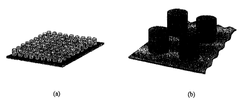
FIGURE 1 shows a schematic diagram of gel posts of 1 mm diameter and 1.1 mm in
height
with (a) the 9x9 array of gel posts and (b) an enlarged diagram of four posts
in the array;
FIGURE 2 shows multi-step preparation of multi-primed gel array;
FIGURE 3 shows a schematic diagram of the instrument used for performing PCR
and
MCA;
FIGURE 4 shows real-time PCR in gel posts arrays with (a) raw fluorescence
data obtained
by CCD image, (b) processed data as contemplated herein and (c) Cp values
obtained for each
post;
CA 02809729 2013-02-27
WO 2012/027832 PCT/CA2011/000989
-5-
FIGURE 5 shows product detection in gel post arrays using melting point
analysis with (a)
melting curves of BK virus (BKV) amplicons in gel posts represented in Fig. 4,
(b) the
negative derivative of fluorescence versus temperature showing the melting
point of the PCR
product and (c) part of the sequence of the product;
FIGURE 6 shows PCR and MCA results for Herpes Simplex virus 1 in a genital
swab made
with primers sequestered in gel-filled wells with (a) raw and (b) normalized
PCR, and MCA
charts with positives coloured blue and negative green based upon (c)
fluorescence and (d)
derivative excitation;
FIGURE 7 shows a comparison of BKV DNA PCR in a 2.8% polyacrylamide gels
performed
in a Lightcycler (a-c) or gel posts (d-f) in particular (a) Lightcylcer real-
time PCR
intensity (b) Lightcylcer Cp values versus logarithm of DNA quantity per 0.85
mL reaction,
(c) size confirmation of the Lightcylcer amplified products in a vertical
polyacrylamide gel,
(d) gel post real-time PCR intensity (e) gel post Cp values versus logarithm
of DNA quantity
per 0.85 1AL reaction, (f) size confirmation of the gel post amplified
products in a vertical
polyacrylamide gel;
FIGURE 8 shows amplification of a target sequence from human genomic DNA by
PCR in
2.8% polyacrylamide gel posts with (a) real-time PCR curves for HPA1, (b)
melting curve
analysis for HPA1, (c) real-time PCR curves for FGFR2 and (d) melting curve
analysis for
FGFR2;
FIGURE 9 shows an (a) amplified PCR product from BKV template applied in
checkerboard
pattern with isolator; (b) qPCR and (c) MCA analysis of positive and negative
posts
demonstrating a clear separation of curves and (d) an 8% polyacrylamide gel
electrophoresis
of DNA in posts showing the specific (111 base pair) and non-specific PCR
products;
FIGURE 10 shows the effect of polymer component of isolator on cross-
contamination
between hydrogel posts using (a) 1% linear polyacrylamide (b) 1% Dextrane 500
(c) 1%
Ficoll 400 and (d) 1% polyethyleneglycol Carbowax 8000;
CA 02809729 2013-02-27
WO 2012/027832 PCT/CA2011/000989
-6-
FIGURE 11 shows PCR and MCA results of BKV DNA amplification with different
product sizes for a multiple primer post array with (a) raw and (b) normalized
PCR, (c) Cp
values and MCA charts with (d) 1 C and (e) 0.25 C resolution.
DETAILED DESCRIPTION OF THE PRESENT INVENTION
The novel method and system described herein provides for the performance of
PCR or other
amplification or gene detection method in a gel medium less than I lit in
volume, obtaining
real-time data in situ by detecting the fluorescence of DNA in the presence of
an intercalating
dye or other means of product or amplicon detection. By performing replicate
PCRs in
multiple gel posts, statistical data to confirm a result can be obtained. The
method of the
.. present invention can be implemented for detection in the same sample of
multiple nucleic
acids, mutations/polymorphisms contained within a heterogeneous nucleic acid
population, or
multiple organisms, pathogens, bacteria or viruses within a single sample. The
use of multiple
different primers added to different gel posts allows a complete set of
simultaneous tests, for
example in clinical sample assays, with requisite positive and negative
controls on the same
gel post array to validate each test run. The multiple posts comprising a
polyacrylamide, or
other cross-linked polymer, hydrogel post array allow different posts to have
different
content, for example loading of differing oligonucleotide primers. Since
nucleic acids are
intended to be analyzed as a single specimen, the individual posts may contain
different pairs
of primers so they can amplify multiple sequences from the genome within a
single post array
without significant cross-contamination. Therefore the art is in need of a
method and
apparatus to perform multiple, essentially independent, nucleic acid
amplification or detection
reactions within a hydrogel post array, in which at least two different primer
sets are present
within the hydrogel post array.
As used herein, an "isolator" refers to a viscous component, as further
described herein,
mixed with at least one component intended to vary between posts within the
hydrogel post
array.
CA 02809729 2013-02-27
WO 2012/027832
PCT/CA2011/000989
-7-
The system described herein is designed to facilitate performance of
diagnostic tests in
parallel on the same sample, using different posts in the same array. A non-
limiting example
of the utility of this platform is testing of patients as the patient presents
in the clinic, for more
rapid results, rather than transport of patient samples to a distant or
centralized laboratory.
This, advantageously, allows for samples to be tested individually, as needed,
rather than
being pooled or transported to distant laboratories for processing. The
ability to acquire real-
time PCR and MCA using the method and system of the present invention expands
the use of
this technique to applications such as isothermal amplification, allele-
specific PCR or
asymmetric PCR for mutation scanning and genotyping performed with unlabelled
probes.
The novel in-gel PCR system of the present invention can perform PCR, melt
curve analysis
and real time quantitative PCR, with an output that compares favourably with
conventional
systems representing a "gold standard". Templates from a viral genome and from
human
genomic DNA are successfully amplified in the gel posts, with BK virus
("BKV"), by way of
non-limiting example, readily detected in unprocessed sub-microliter volumes
of urine from
patients with BK viruria. Further, it is contemplated by the present invention
that both
processed and unprocessed clinical samples other than urine may be used with
the present
method and system, including but not limited to, serum, plasma, whole blood,
sputum,
mucous, aspirates, debrided tissue, scrapings and lymphatic fluid. Further,
the present
invention is not limited to use with only clinical samples from humans or
animals, as the
systems and methods described herein may use any sample which may contain a
template for
the amplification or detection as contemplated herein such as genetic or
molecular
characterization of bacteria, plant, mould, fungus or other lower-organism.
The present
invention contemplates use of methods for detecting a gene or transcript other
than PCR and
one skilled in the art would be aware of the variations of PCR and other gene
or transcript
detection systems.
The present invention provides a method of performing real-time PCR in gels
with MCA in
an array of cylindrical shaped self-standing gel posts (-0.64 - 0.86 L per
post). In a
preferred embodiment the PCR and post-PCR analysis of the resulting amplified
nucleic acid
CA 02809729 2013-02-27
WO 2012/027832
PCT/CA2011/000989
-8-
(if any) was performed in microfluidic volumes utilizing, in one embodiment, a
9x9 pattern of
posts (Fig. 1). An inexpensive prototype heating device with a Peltier element
was used for
performing PCR and MCA, a diode laser for excitation of fluorescence, and
detection optics
containing a CCD, all of which controlled by a micro-controller. As well, the
present
invention provides the novel and desirable performance of in-gel amplification
of templates
from genomic DNA ("gDNA"), cDNA or RNA from human, animal, bacteria, plant,
mould,
fungus or other lower-organisms.
The gel of the present invention is contemplated to hydrophilic polymers
forming colloidal
hydrogel matrixes which result in similar mobilities of the nucleic acids of
sizes contemplated
by the present invention as in the specifically described gels herein, by way
of non-limiting
example, polyacrylamide cross-linked with bis-acrylamide and
polyvinylpyrrolidone
("xPVP') cross-linked with PEG-diacrylate resulting from the
photopolymerization of 3.3%
vinylpyrrolidone with 0.7% Polyethyleneglycol-diacrylate.
The posts contemplated by the present invention may be cylindrical, spherical,
conical or any
other shape and dimension, so long as the posts of the array are physically
separated, though
they may be in fluid communication or in a common fluid substrate. The size
and shape of
the posts presented herein are presented as exemplar structures, and it is
contemplated that a
variety of moulds and therefore post shapes are possible. Also contemplated
are inverted
shapes placed on a planar surface, for example wells or depressions comprised
of a hydrogel.
Also contemplated are wells, depressions or capillaries made within a
structure, for example
plastic, glass, metal or other materials, filled with hydrogel.
There are significant disadvantages to the introduction of primers to
individual posts within
the post array through physical means. Primers present in a post forming part
of a post array,
as contemplated by the present invention, must be allowed sufficient time to
diffuse
throughout the hydrogel post prior to the nucleic acid sample to which the
primers are
intended to anneal, coming into contact with the post. As well, the primers
within the post
must be accurately introduced into the post so as to prevent cross
contamination between the
CA 02809729 2013-02-27
WO 2012/027832 PCT/CA2011/000989
-9-
posts forming the post array; which is a challenge given the small volumes and
limited
spacing between posts as contemplated herein. Deposition of the primer mixture
on a mold
prior to introduction of polymer is of limited benefit, as the evaporation of
the primer solution
changes the localized salt concentration which thereafter affects the melting
temperature of
the PCR product and may even adversely impact the ability of the polymerase to
catalyze
PCR. It has been observed that in square arrays the primer solutions deposited
within a planar
mould results in evaporation on the outer edges at a rate faster than the
innermost posts, and
the rate of evaporation is difficult to control. Further, the prior art method
of depositing
primers in a well prior to the addition of polymerization reagents,
disadvantageously resulted
in rapid mixing with the gel when it is added and resulted in extensive cross
contamination.
Addition of primer components to the posts once polymerized is mechanically
intensive and
ensuring delivery of equal molarity of primers is technically challenging.
The present invention contemplates the deposition of primers to a mould, the
deposition
limited to a region consistent with, or internal to, a planned hydrogel post
following a
polymerization step as contemplated herein; wherein the primers are mixed with
a viscous
component for deposition, the viscous component selected from candidates
including, but not
limited to, carbohydrates, polymers or carbohydrate-polymer mixtures; such
that following
evaporation, the primer and viscous component forms a film on the mould which
temporarily
prevents primers from dissolving in the master mix during mould filling and
covering; and
allows polymerization of the hydrogel to occur while the primer and viscous
components
dissolves within the forming hydrogel post, and which will not interfere with
the detection
process used for the nucleic acids, as further described herein. As the
primers are diffusing
from within the polymerizing hydrogel post, the time needed for photo-
polymerization is
sufficient for the primers to distribute essentially evenly throughout the
post. Following
detachment of the mould and submersion of the array into oil or other medium,
as
contemplated herein, the posts have primers present within the individual
posts. These
primers are isolated from adjacent posts and do not cross-contaminate each
other, enabling
multiple amplification reactions using different nucleic acid primers or
templates to occur on
CA 02809729 2013-02-27
WO 2012/027832
PCT/CA2011/000989
-10-
the same gel post array, including some posts that lack any primer at all
(negative control).
Although in one embodiment every post could harbour a different set of
primers, in practice,
other embodiments would be groups of posts that all have the same primer set
(replicates)
and/or primer sets arranged in a "checkerboard" pattern wherein each region of
the
checkerboard has a different set of primers to amplify a different template
from the same
sample. The viscous component and optional variable elements included therein,
are referred
to as the "isolator".
The present invention contemplates the use of various mono- and disaccharides
as the viscous
component, though in the preferred embodiment the viscous component is
sucrose. Ones
skilled in the art will recognize that a variety of agents, soluble in water
and other than
saccharides, are capable of being used as the viscous components, selected for
their ability to
temporally retard primers from dissolving into the unpolymerized hydrogel of
the present
invention on filling and covering, and further selected based upon their
interaction with PCR
and polymerization. Optionally, visible dyes may be added with the viscous
component so as
to allow visualization of evaporation and restriction of the associated primer
to particular
posts within the post array. In a preferred embodiment the viscous component
has a pH
greater than 7.0, so as to inhibit annealing of the primers during the
evaporation; although
other pH values are contemplated by the present invention, as well as the
inclusion of
components which inhibit annealing of the primers during evaporation.
Example 1: Gel Polymerization
The polymerization of acrylamide gel for gel PCR can be initiated either by a
photochemical
method or by using peroxide. Adding ammonium persulfate (APS) as the initiator
peroxide is
the widely used method (Sambrook, J. & Russel, D. W.; Molecular Cloning, 3rd
Edition ed.;
CSHL Press, 2001). For photochemical polymerization, `azobis' (2,2'-azobis(2-
methyl-N-(2-
hydroxyethyl) propionamide)) or riboflavin or Methylene Blue is added to the
gel mix and the
photochemical reaction is started by exposing the gel mix to ultraviolet
light. It was noted that
the polymerization initiator, APS, inactivated or inhibited the fluorescent
intercalative dyes
CA 02809729 2013-02-27
WO 2012/027832 PCT/CA2011/000989
-11-
such as SYBR Green I and LC Green Plus that are needed for subsequent product
detection
by intercalative fluorescence, precluding addition of dye prior to the
polymerization if the
APS is used as the polymerization initiator. To circumvent this problem the
gel posts were
made by photo-polymerizing the PCR reaction mix with LC Green Plus or SYBR
Green, with
or without template DNA, along with the acrylamide gel reagents in a glass
mold having, for
example, a 9x9 array of wells (Fig. 1). An alternate embodiment is to
introduce the
intercalating dye or other agent after completion of PCR or other
amplification reaction. The
wells were then sealed with a cover slip treated to promote gel adhesion. Once
gel
polymerization had occurred, the cover slip was detached from the mold along
with the array
of gel posts, and immersed in mineral oil to minimize evaporation, as
described below.
Example 2: Mould & Cover slip preparation
Fig. 2 shows a summary of the steps associated with the preparation of the
multi-primer
hydrogel post array. Wells are filled with primer-isolator mix, Fig. 2(a);
dried, Fig. 2(b); the
PCR-polymerization mix is added and the mould covered with a cover slip, Fig
2(c); after
photo-polymerization, Fig 2(d); the cover slip with hydrogel posts is detached
from the
mould, Fig. 2(e). The mould, approximately 20 mm x 20 mm, is made with a 1.1
mm thick
glass microscope slide permanently bonded to another 1.1 mm thick microscope
slide with a 9
x 9 or 6x4 array of holes of cylindrical or conical shape 1 or 2 mm in
diameter, although it is
contemplated that other shapes of arrays and posts are also possible. Post
arrays may be
removed from the mould for use as reaction vessel, or in an alternate
embodiment may remain
within the mould or other support, for an enclosed reaction. Polyacrylamide
gels of 2.8% to
12% are contemplated, with 2.8% being the softest gels reported for in-gel
PCR.
To prepare the surface of the mould so that it would not adhere to the gels, a
thin layer of
Sigmacote (Sigma, St. Louis, MO, cat# SL2) was spread onto the surface of the
mould and
left to dry. The mould was then washed with n-heptane (Applied Bio Systems,
Foster City,
CA, cat # 400079) and blown dry with air. In contrast, the surface of the
cover slips (22 mm x
22 mm, Fisher, Fair Lawn, NJ, cat#12-54B) were treated to enhance adherence to
the gel by
CA 02809729 2013-02-27
WO 2012/027832 PCT/CA2011/000989
-12-
immersing them in a mixture of 40 mL of 95% ethanol, 1 mL of 100% acetic acid
(Fluka,
Buchs, cat# 45725), 8.9 mL of water, 100 L of 3-(trimethoxysilyppropyl
methacrylate
(Sigma, cat# 440159) for 1 hour followed by washing with isopropanol (2-
propanol). After
preparing the isolator, and casting a gel, the mould can be washed and reused
for subsequent
gel casting or the mould can remain permanently in contact with the gel. In
another
embodiment, the gel post array may be fully enclosed with a port for
introduction of template
and/or other reagents.
Example 3: PCR/MCA Process, BKV PCR
In order to observe the characteristics of polymers and their effect on
nucleic acid detection or
amplification within hydrogel post arrays, hydrogel post arrays were prepared
absent the use
of the isolator contemplated by the present invention. After depositing
primers in the isolator,
the gel can be polymerized with or without template DNA included in the
polymerization
mixture. In the latter case, the DNA can be added atop the gel posts where it
diffuses into the
gel before PCR is performed. One hundred L PCR gel mix contained 47 I, PCR
reagents,10 L gel reagents and 43 1, water. The 474 PCR reagents were: 20 L
of 5 x
PCR buffer (333 mM tris-sulphate, pH 8.6, 83 mM (N}14)2SO4 (Sigma); and 40%
sucrose
(Sigma)), 4 I, of 50 mM MgCl2 (Fluka), 2 L of -10mM [dNTPs] (Sigma), 2 111,
of 1%
bovine serum albumin (Sigma), 2 p.IL of 10 M primer solution (Integrated DNA
technologies, San Diego, CA) for each of 2 primers to produce 100 base pair
("bp") product, 2
1, BKV template DNA either before or after polymerization, 10 1., of 10xLC
Green Plus
(Idaho Technology Inc., Salt Lake City, Utah) and 3 4 of Taq polymerase (20
units/AL). The
10 4 of gel reagents were: 7 L of a 40% acrylamide (Sigma, cat # A9099) + 4%
bis-
acrylamide aqueous solution (N,N-methylene bisacrylamide, BioRad, Hercules,
CA, cat#
BA05-1610201), 2 AL of 3% azobis (Wako, Richmond, cat# VA-086), and 1 4 of 10%
TEMED (N,N,N',N1-tetramethylethylenediamine, Sigma, cat# T7024). This mixture
was
degassed in vacuum and pipetted into the wells in the mold. Once all the wells
were full, a 22
mm x 22 mm cover slip treated, as noted above, for adherence to the acrylamide
was slipped
CA 02809729 2013-02-27
WO 2012/027832 PCT/CA2011/000989
-13-
atop the wells. The isolator prevents cross contamination of different primer
sets during this
step. The mold with the cover slip atop was then exposed to the 405 nm laser (-
4 mW/cm2 on
the posts) for 25 mm in order to photo-polymerize the acrylamide mix. The
cover slip with the
attached posts was then slowly lifted from the mould, immediately immersed in
mineral oil
(Sigma, cat# M5904) in a shallow anodized aluminum 23 mm x 23 mm pan (posts
facing up),
and placed on the Peltier element. Thermal cycling was performed with an
initial denaturation
of 30s at 94 C followed by 50 cycles of denaturation at 94 C for 15s,
annealing at 52 C for
30s, and extension at 72 C for 30s, and ending with an extension step of 72 C
for 60s. After
completion of PCR, MCA was performed. To determine the threshold for BKV
amplification,
BKV PCRs were performed with 34 to 8640 BKV copies/post. Overall, a total of
52/52
independent experiments to amplify BKV were successfully performed on gel
posts,
confirming reproducibility of the method.
In order to show that PCR can be performed with unprocessed samples, PCR was
performed
with 1.5 L, of raw urine added to the PCR reaction mix prior to the
polymerization. All the
PCR parameters are similar to the BKV DNA PCR other than the PCR cycle number
was
reduced to 35.
For addition of template after polymerization of gel posts, a similar PCR gel
mix (as above)
was made without BKV DNA and polymerized. After the gel was detached from the
mould, a
14 I, BKV template (2.86 x10⁷ copies/mL) was added atop the gel posts and the
DNA
allowed to diffuse for 30 min in a covered Petri-dish before performing PCR
with the same
thermal cycle conditions as above. If the DNA added atop the gel was uniformly
absorbed, the
amount of template DNA was 4,900 BKV copies/post. Real time quantitative PCR
confirmed
that the same template copy number was detected for template polymerized
within the gel or
added atop the gel. In order to study the size limitation of the product that
could be amplified
in a given gel concentration, we have also performed BKV PCR (17,280 BKV
copies per
post) with a series of different primers (Table 1), with the template DNA
polymerized in the
gels as indicated above.
CA 02809729 2013-02-27
WO 2012/027832 PCT/CA2011/000989
-14-
Table 1: Primer sequences for BKV and HPA1 amplification by PCR
Primer SEQ ID Product
description length Sequence
(bp)
BKV reverse SEQ ID NO 1 5'-aaacaccctaacctcttctac-3'
BKV forward SEQ ID NO 2 100 5'-ttcctttttgctaagtgacc-3'
SEQ ID NO 3 150 5'-tattttaagatccgcctga-3'
SEQ ID NO 4 200 5'-gcctgtttactaacagctctg-3'
SEQ ID NO 5 250 5'-gcctctttgtaaagctgatag-3'
SEQ ID NO 6 300 5'-catgtgaccaacacagctac-3'
SEQ ID NO 7 350 5'-ctaggtattttgggactttca-3'
SEQ ID NO 8 400 5'-tgcttatccagttgagtgc-3'
SEQ ID NO 9 450 5'-ccagtcccaggtaatgaatac-3'
SEQ ID NO 10 500 5'-gaattacaggtcaaagtaccc-3'
SEQ ID NO 11 600 5-gtgcatgagcatggtgga-3'
SEQ ID NO 12 800 5'-aagctaagtgctgaaaatgac-3'
SEQ ID NO13 1000 5'-cccaac,caaaagaaaagg-3'
HPA1 reverse , SEQ ID N014 5'-cacagcgaggtgagcc-3'
HPA1 forward SEQ ID NO 15 42 5'-ctcctgtcttacaggccc-3'
FGFR reverse SEQ ID NO 16 5'-gctgacttctatttatataacttcaagc-3'
FGFR forward SEQ ID NO 17 5'-cagaagtttttgagagtggcatgatg-3'
HSV1 reverse SEQ ID NO 18 5'-cgccggcggatacgaagacg-3'
HSV1 forward SEQ ID NO 19 174 5'-cgtcgcgggttggccacata-3'
Example 4: PCRJMCA from a clinical sample in gel-filled wells
A glass mould 6x4 was prepared as described in example 2, filled with 0.2
1.t.M HSV1 primers
(Table 1) in 8% Trehalose and dried for 1 hr. Then PCR gel mix was added into
the wells.
One hundred 1.1.1., PCR gel mix contained 41 p.L PCR reagents,13 111, gel
reagents and 43 !IL
water. The 47 L, PCR reagents were: 20 1..tL of 5 x PCR buffer (333 mM tris-
sulphate, pH
8.6, 83 mM (NH.4)2SO4 and 40% sucrose, 4 1.11, of 50 mM MgCl2, 2 tiL of -10mM
dNTPs .2
pi of 1% BSA, 10 1., of 10xLC Green Plus and 3 p.L of Taq polymerase (20
units/A). The
10 p,L of gel reagents were: 10 tit of a 40% acrylamide + 4% bis-acrylamide
aqueous, 2 gL
CA 02809729 2013-02-27
WO 2012/027832 PCT/CA2011/000989
-15-
of 3% azobis and 1 1tL of 10% TEMED. The reagent suppliers are same as in
example 3. A
cover slip was then slid over the mould and acrylamide was photo-polymerized
for 22 min
under 360 nm UV lamp (UVG L25, UVP, Upland, CA). The cover slip was then
removed and
44 of a clinical sample arising from immersion of a genital swab in Universal
transport
medium (Copan Diagnostics Inc. Murrieta CA) diluted with 21 ill of 1xPCR
buffer was
applied on the top three rows of the hydrogel well array (one half of the
total number of
hydro gel filled wells) for 10 min. A multiplicity of sample treatment
protocols for application
to gel posts can be envisaged by one skilled in the art who would recognize
that the final
volume depends on the number of gel posts to which the sample is applied. The
sample was
removed and the mould was then immersed in mineral oil in aluminum pan.
Thermal cycling
was performed with an initial denaturation of 90s at 96 C followed by 35
cycles of
denaturation at 94 C for 30s, annealing at 63 C for 40s, and extension at 72 C
for 35s, and
ending with an extension step of 72 C for 120s. After completion of PCR (Fig.6
(a) and Fig.
6(b)), MCA was performed (Fig.6 (c) and Fig. 6(d)). Negative samples
demonstrate
separation from positive within Fig. 6 as the lower, light-gray, lines within
the individual
graphs and positive samples represented by the darker lines above.
Example 5: PCR/MCA Process, Genomic DNA PCR
Extending the principles from Example 3, similar PCRs were performed with
purified gDNA
added to the gel before or after the polymerization. Overall, gDNA has been
successfully
amplified in gel posts for 27/27 independent experiments. For the PCR
performed with
gDNA template polymerized in the gel, a 42 bp product from human HPA1 (human
platelet
antigen 1) was amplified. Except for the template and oligonucleotide primers
(as listed in
Table 1), the PCR reaction mix was similar to that for the BKV PCR. 225 ng of
gDNA was
added to the 100 tit mix (-2.2 ng per post). PCR thermal cycling conditions
were as indicated
for the BKV PCR.
For the PCR performed with gDNA added atop the polymerized gel, a 71 bp
product from the
FGFR2 gene from human gDNA was amplified. Primers used for FGRF2 amplification
were
CA 02809729 2013-02-27
WO 2012/027832 PCT/CA2011/000989
-16-
forward primer SEQ ID No. 16(5' "CAGAAGTTTTTGAGAGTGGCATGATG") and
reverse primer SEQ ID NO 17(5' "GCTGACTTCTATTTATATAACTTCAAGC"). Fourteen
1AL of gDNA (30 ng/4) was pipetted onto the whole array of posts and was left
in a covered
Petri-dish for 30 min to allow diffusion of gDNA into the gel. If all the gDNA
was uniformly
absorbed, a DNA amount of ¨5 ng/post is predicted.
Example 6: PCR/MCA Instrumentation
An inexpensive prototype instrument (shown in Fig. 3) is used to perform the
PCR reaction in
the gel posts. This instrument uses Motorola 68332 microprocessor 307 to
control Peltier
element 304 (XLT2398-01L, Marlow Industries, Dallas, TX) to perform heating
and cooling
for PCR and MCA, where Peltier element 304 is placed in thermal communication
with
hygrogel array 310 and heatsink 305. A charge-coupled device ("CCD") camera
301 (Deep
Sky Imager, Meade, Irvine, CA) is mounted above Peltier element 304 as well as
hydrogel
array 310. 65 mW 405 nm laser diode 306 (DL-7146-101S, Sanyo) is mounted at a
70 degree
angle to horizontal for a fluorescence excitation source. Laser diode 306
delivers an average
of 32 IAW of excitation power to each post within hydrogel array 310. 50 nm
wide band-pass
interference filter centered at 510 nm 302 (BP510/50, Chroma Technology,
Bellows Falls,
VT) is mounted in front of the camera to attenuate excitation light. Biconvex
lens 303
(KBX046, Newport, effective focal length 25.4 mm) is mounted between filter
302 and
hydrogel array 310. Camera parameters such as the exposure time, light and
dark levels are
set by the user on PC compu1er308, in electronic communication with
microprocessor 307.
During PCR, once the extension temperature is reached, laser diode 306 is
switched on and a
fluorescent image of the gel posts is taken by CCD camera 301 and stored by
computer 308.
During the MCA, laser diode 306 is left on continuously and an image is taken
by CCD
camera 301 and stored by computer 308 at each degree from 50 C to 95 C, once
the chip has
been stabilized at a particular temperature. The system is calibrated by
placing calibrated
thermocouple 309 (5TC-TT-K-40-36, Omega Engineering Inc., Stamford, CT)
between the
hydrogel posts within hydrogel array 310, under the oil. The settings on the
system are then
correlated to the observed temperatures of the calibrated thermocouple.
Microprocessor 307 is
CA 02809729 2013-02-27
WO 2012/027832
PCT/CA2011/000989
-17-
in electronic communication with, and controls, Peltier element 304,
temperature sensor 309
and laser diode 306.
Example 7: PCR and Melting Curve Analysis
The CCD images acquired at the extension step of each PCR cycle (total of 50
images) were
analysed with ImageJ software (National Institutes of Health, USA) using the
MicroArray
Plug-in (Dr. Robert Dougherty, OptiNav Inc., Redmond, WA) that can be used to
plot the
cycle number vs. the fluorescence intensity of each post. Even though the
mould disclosed
herein creates a 9 x 9 array of posts, optical limitations of the CCD assembly
allow image
acquisition for only a 6x8 array. In order to determine the efficiency of the
PCR, most
commercial real-time quantitative PCR instruments embed some proprietary
version of data
processing in their software. All reported methods characterize the real-time
PCR curves by
applying curve fitting and determining the threshold values where the
fluorescence of the
PCR begins to rise above the background signal and the DNA copy numbers can be
seen to
increase exponentially.
Therefore a sigmoid was fitted to the real-time PCR curves in order to find
the exponential
region and to find the threshold value, termed the "crossing point" (CP). A
Linear Regression
of Efficiency or LRE method (Rutledge, R. G. & Stewart, D. BMC Molecular
Biology 2008,
9) modified with a linear baseline correction (Rebrikov, D. V. & Trofimov, D.
Y. Applied
Biochemistry and Microbiology 2006, 42, 455-463) to fit the sigmoid.
With LRE, the fluorescence of the DNA, Fe at cycle c of the PCR can be written
as
Max
+ F + cF
Fo 1XE mar -F Formula!
where Fo, Froax, Flei and FK are the fluorescence values for the initial
reaction, endpoint
reaction, constant background and variable background, and 443X is the maximum
amplification efficiency. A linear baseline to the equation was used to
facilitate baseline
CA 02809729 2013-02-27
WO 2012/027832 PCT/CA2011/000989
-18-
subtraction with the last two terms of the equation, F8 and cFK. The maximum
of the second
derivative of the sigmoid is determined to calculate the Cp value,
representing the cycle
number at which the fluorescence has risen above the background level and the
exponential
growth of the PCR is at a maximum.
The algorithm of Formula 1, was implemented into computer code using visual
basic for
applications 6.3 in Microsoft Excel, which receives the fluorescence
intensities obtained from
the text file output by the Image.1 software and returns multiple plots
including the raw, fitted,
and normalized data. Cp values for each PCR reaction are also calculated for
each post. Fig.
4(a) shows real-time data (experimental data points connected by interpolated
lines) for 36
posts that were obtained after PCR performed with 3,456 starting copies of BKV
DNA
template per post in a 2.8% polyacrylamide gel. Insets in Fig. 4(a) show CCD
images of the
gel array at the 1st cycle, 30th cycle, and 50th cycle. These results were
confirmed in more than
10 independent experiments and no fluorescence above background was detected
in the
negative controls Fig. 4(b) and Fig. 4(c) show the plots produced by the
algorithm and the Cp
values for each post respectively.
There is spatial variation in the illumination of the post array due to the
oblique incident angle
and intentional optical diffusion of the laser. As a result, fluorescence
excitation is not
uniform on all posts, and thus each real-time PCR curve starts at a different
intensity level as
seen in Fig. 4(a). These background variations are removed by data processing
to produce the
normalised curves of Fig. 4(b). Considerable background light between posts is
observed, as
shown in the inset post array image for the 50th cycle in Fig. 4(a). This
background is due to
a thin film of gel that remains between the posts as the gel post array is
assembled, and where
PCR also occurs. One skilled in the art would recognize that modification of
assembly
protocols would remove or reduce this thin film, and it is contemplated that
the present
invention also encompasses such modified assembly protocols. Despite the
presence of the
thin gel layer, fluorescence data is largely independent of the background as
they result from
the summation of pixels entirely within each post. As disclosed herein, the
present invention
does not suffer from "cross-talk" between posts.
CA 02809729 2013-02-27
WO 2012/027832 PCT/CA2011/000989
-19-
Melting of the DNA was performed immediately after the PCR was completed. The
melting
curves were obtained by measuring the fluorescence in the CCD images obtained
at each
degree from 50 C to 95 C as seen in Fig. 5(a). The negative derivative of
the fluorescence
with respect to the temperature was plotted in Fig. 5(b) and allowed the
melting temperature
of the PCR products (T. ) to be determined as the temperature at the peak 31.
The melting
temperature for BKV amplicons (average T. 1 a for all 36 traces) was 82.6
0.4 C. The
sequence of BKV PCR product was confirmed by sequencing the DNA from one post.
Part of
the sequence is shown in Fig. 5(c) with sequencing performed with ABI 3130x1
DNA
capillary analysis system (Applied Biosystems, Foster City, CA). As for the
real-time PCR
traces in Fig. 4(a), the melting curve baselines of Fig. 5(a) are influenced
by the
heterogeneous laser illumination; this bias is removed through the data
differentiation used to
produce Fig. 5(b). Also in keeping with Fig. 4(a), the inset image for 75 C
shows
considerable background fluorescence, owing to the thin layer of gel that
remains between
posts. Insets in (a) show the CCD images of the gel array at 75 C and 85 C.
These results
were confirmed in more than 10 independent experiments.
The results shown in the Fig. 4 and Fig. 5 were acquired with the gels
polymerized with the
BKV DNA template inside. However, if this technology is to be applied to real-
world medical
diagnostics, adding clinical samples to the pre-cast gel is likely to be a
better approach. PCR
with a BKV DNA template that was allowed to diffuse into a pre-cast gel matrix
was
performed and real-time PCR curves obtained, as shown in Fig. 7(a). This
confirms that
exogenous template DNA can successfully enter the gel and interact with the
embedded PCR
components. The melting curve analysis data is shown in Fig. 7(b); for the
experiment shown,
the average melting temperature was 82.8 0.6 C.
Example 8: Effect of the gel concentration on the PCR product size
In order to study the limitations of PCR product lengths in polyacrylamide
gels, a series of
BKV products with lengths from 100 to 1000 bp in 5 different gel
concentrations were
amplified absent isolator, using the method described in Example 3, with
template DNA
.1
CA 02809729 2013-02-27
WO 2012/027832
PCT/CA2011/000989
-20-
added prior to polymerization and primers added before polymerization using
the isolator
method described herein. Table 2 shows PCR amplification of different lengths
of BKV
template in different polyacrylamide gel concentrations, with a (+) sign
indicating that PCR
product was detected. Thirteen different primer sets were used to amplify
different sized
segments of a BKV template, using 5-6 different primer sets per gel post array
(see Table 1).
Primers were added to the moulds prior to polymerization (isolator method).
Results were
confirmed with at least two experiments for each primer set and the sizes of
the PCR products
were confirmed by running vertical in situ gel electrophoresis on each post.
Each array
included several different primer sets, deposited by the isolator method, for
distinct sets of gel
posts, demonstrating simultaneous multiparameter testing. The results
demonstrate that the
gel limits the size of the product that can be amplified. As the gel
concentration increases, the
maximum product size that can be amplified decreases suggesting that the
smaller pore size of
harder, high concentration gels restricts movement of the larger reagent
molecules (DNA
template, polymerase etc.) inside the gel as compared to their movement in
lower
concentration, softer gels. The size limits shown in that table are
appropriate for PCR
annealing and extension times of 30s as postulated in Example 3.
Table 2: BKV template amplification length, gel concentration and cross-talk
controls.
Concentration of Polyacrylamide
Size of
4% 6% 8% 10% 12%
Amplicon (bp)
100 + +
150 + +
200
250
300
CA 02809729 2013-02-27
WO 2012/927832
PCT/CA2011/000989
-21-
350
400
450
500
550
600
800
1000
In order to show that the primers do not diffuse from one post to another, a
separate
experiment was performed in which a single primer set was added to some but
not all wells
prior to the addition of the gel polymerization mix as described in Example 4
to create a
checkerboard of adjacent positive and negative controls. Table 3 shows
positive posts have
PCR amplification of 100bp PCR fragment in 6% polyacrylamide gel while
negative posts
lacked the primer to allow amplification. The lack of amplification observed
confirms that
cross-talk is suppressed. No product was obtained for posts lacking primers,
indicating that
when using the isolator method described herein, cross-contamination by
primers does not
occur. Diffusion of PCR components between posts was not detected.
Table 3: Cross-talk controls via checkerboard of alternating positive and
negative posts.
posts with primers (100bp) posts without primers
Example 9: Quantitative PCR
In order to characterize quantitative real-time PCR in gel posts, different
amounts of BKV
CA 02809729 2013-02-27
WO 2012/027832 PCT/CA2011/000989
-22-
DNA were tested under the same PCR conditions to amplify a product of 100 bp
in 2.8%
polyacrylatnide gel posts. For comparison, conventional real-time PCR with the
same
template was carried out in the Lightcycler , an instrument that is routinely
used for melt
curve analysis in clinical diagnostic laboratories and provides a clinically
relevant "gold
standard". For the Lightcycler PCR, PCR reaction/gel mixes were polymerized
in capillaries
in order to mimic the PCR in the gel posts and held a total volume of 0.64 -
0.84 1AL per
reaction, similar to gel posts, though smaller volumes are also contemplated.
Fig. 7(a-c)
shows real-time PCR data generated by the Lightcycler , relationship of Cp
values versus log
[DNA]mittal and the confirmation of the product size by vertical gel
electrophoresis. The
analogous results of Fig. 7(d-f) obtained with the in-gel post PCRs mirror
those of Fig. 7(a-c)
from the Lightcycler . Each post was picked up individually and placed above
the gel before
running electrophoresis as shown in Fig. 7(f), with a 100 bp DNA ladder shown
in the middle
in Fig 7(1) and on the left in Fig. 7(c). The results below confirm that melt
curve analysis of
PCR in gel posts matches that from gold standard testing.
Fig. 7(b) and Fig. 7(e) show that, as expected, the Cp values decrease
linearly with the
logarithm of increasing template DNA copy number for the Lightcycler and gel
post array,
respectively, and that the relationship is comparable in the two systems
(within ¨1 cycle).
The melting temperature of the products in the Lightcycler is 81.5 C which
agrees with the
gel posts value of 82.6 C. Both real-time PCR and MCA validate the PCR
conditions in gel
posts. The inset in Fig. 7(f) is an enlarged section of the gel band, showing
individual bands
from each gel post. The samples loaded to electrophoresis lanes shown in Fig.
7(1) were from
PCR performed with an initial 3456 BKV DNA templates per post.
Example 10: Genomic DNA PCR
Human gDNA is made of 3 billion base pairs of DNA, as compared to viral DNA,
or plasmid
DNAs that are only a few thousand to hundreds of thousands of base pairs in
size. During
PCR, there is a great deal of heterogeneous non-target DNA present in the long
gDNA
compared to the uniformity of short plasmid DNA, suggesting that the
efficiency of the
CA 02809729 2013-02-27
WO 2012/027832 PCT/CA2011/000989
-23-
gDNA PCR is less than that of plasmid DNA. The prior art with respect to gel
PCRs used
plasmid DNA or cDNA as the template but not gDNA. Two gDNA PCR were undertaken
in
2.8% polyacrylamide gel, one with the gDNA polymerized in the gel and one with
the gDNA
added after polymerization of the gel.
gDNA was subjected to PCR to amplify a 42 bp product containing a known SNP
from the
human HPA1 gene in the gDNA template polymerized inside the gel, using SEQ ID
NOs 14
and 15 as primers; as otherwise shown in Table 1. The template was chosen in
anticipation of
future genotyping with the gel posts using e.g. allele specific PCR as
previously shown. The
processed real-time PCR curves and the melting curve analysis data are shown
in Fig. 8(a)
and Fig. 8(b) respectively. The amount of genomic DNA in the HPA1 PCR is ¨ 2.2
ng per
post. PCR was then performed with gDNA template (-5 ng/post) added after the
gel
polymerization, the gDNA allowed to diffuse into the gel matrix. For the
latter approach, the
FGFR2 gene from human genomic DNA, with amplification of a 71 bp product also
containing a known SNP using SEQ ID NOs 16 and 17 as primers. Fig. 8(c) shows
the
processed real-time PCR curves while Fig. 8(d) shows the melting curve data
for the FGFR2
PCR. Both HPA1 and FGFR2 product sizes were confirmed by vertical gel
electrophoresis.
This is the first use of gDNA for gel PCR, where gDNA is introduced to the gel
mix either
prior to or after the polymerization.
Example 11: Primers incorporated into a post array in checkerboard pattern.
A hydrogel post array was prepared in checkerboard pattern using an isolator
comprised of
8% sucrose, 1% Dextran T500, 13 mM Tris-base, 0.05% NP-40, 0.05% Tween20 and
0.2ILM
of each primer and BKV DNA (-3,000 molecules per post) or no DNA. The isolator
mixture
has a pH of 10.6, which advantageously inhibits primer annealing during the
evaporation of
the isolator, Fig.2(a) and Fig 2(b), Mould heights of 1.1nun were used and
following
polymerization, Fig.2(c) and Fig. 2(d), detachment from the mould, Fig. 2(e),
and immersion
into oil; PCR (Fig. 8(a) and Fig. 8(b)) was undertaken with monitoring of the
PCR through
melting curve analysis (Fig. 8(c)). A vertical 8% polyacrylamide gel
electrophoresis (Fig.
CA 02809729 2013-02-27
WO 2012/027832 PCT/CA2011/000989
-24-
8(d)) demonstrated specific PCR product in positive posts and variations of
primer dimer in
no-template posts.
Example 12: Alternate polymers for isolator mixture.
Alternate polymers were compared in a preparation of 1% polymer with
8%sucrose, with
thinner molds designed to result in hydrogel posts of 0.5 mm as opposed to the
1.1mm used in
other experiments disclosed herein. Fig. 9 shows a comparison of four
hydrophilic PCR-
compatible polymers, linear polyacrylamide (Fig. 9(a)), Dextran 1500 (Fig.
9(b)), Ficoll 400
(Fig. 9(c)) and polyethylene glycol Carbowax 8000 (Fig. 9(d)); and visually
demonstrates
their ability to temporally prevent dissolving of the evaporated isolator
solution.
Bromophenol Blue dye (0.05%) was loaded to every other well along with 8%
sucrose and
dried at room temperature, as a visual idicator of the isolator's ability to
temporarily retard the
dissolution of products within the isolator upon addition of the
unpolyrnerized hydrogel.
Visual comparison demonstrated that linear polyacrylamide had a slight
advantage over the
next best polymer, Dextran T500.
Example 13: Multiple primers incorporated into hydrogel posts within a
hydrogel post array
Inclusion of between 3 to 12 different sets of primers in individual posts
comprising a
hydrogel post array were successfully undertaken using an isolator comprised
of 8% sucrose,
1% Dextran T500, 13 mM Tris-base, 0.05% NP-40, 0.05% Tween20 and 0.21.IM of
each
primer and BKV DNA (-3,000 molecules per post) or no DNA. The isolator mixture
has a
pH of 10.6, which advantageously inhibits primer annealing during the
evaporation of the
isolator, Fig.2(a) and Fig 2(b). Mould heights of 1.1mm were used and
following
polymerization, Fig.2(c) and Fig. 2(d), detachment from the mould, Fig. 2(e),
immersion into
oil.
Twelve primer pairs were used spanning product sizes from 150 to 1000 b.p. in
accordance
with the primers listed in Table 1, using BKV as a template, and isolator
deposited as
described herein. Each pair was represented by four posts providing
statistical value to the
CA 02809729 2013-02-27
WO 2012/027832 PCT/CA2011/000989
-25-
experiments and serving redundancy in case posts were disrupted during
detachment of the
mould. The mould was dried for an hour on open air and transferred to the gas
chamber to be
filled with PCR-polymerization mixture and photo-polymerized as described
herein. The
PCR and MCA were performed as described herein, and the images were processed
in ImageJ
.. and Microsoft Excel with the typical results shown in Fig.11. In order to
prove the size of
amplified products the posts were detached from the glass support and placed
on a vertical 8%
polyacrylamide gel, polymerized in 0.5x TBE buffer (20 mM Tris-Borate, 0.5 mM
EDTA).
Electrophoresis extracted PCR products from the posts and separated them
according to their
sizes in the gel.
While particular embodiments of the present invention have been described in
the foregoing,
it is to be understood that other embodiments are possible within the scope of
the invention
and are intended to be included herein. It will be clear to any person skilled
in the art that
modifications of and adjustments to this invention, not shown, are possible
without departing
from the spirit of the invention as demonstrated through the exemplary
embodiments. The
invention is therefore to be considered limited solely by the scope of the
appended claims.